Even more knowledge

Radiation safety is the cornerstone of contemporary pediatric dental imaging. Digital sensors emit up to 90% less ionizing radiation than traditional film, allowing clinicians to follow the ALARA principle while still acquiring diagnostically useful images. Current low‑dose cone‑beam CT protocols further limit exposure, often matching the dose of a few intra‑oral bitewings.
Digital versus film also drives efficiency. Instant image preview lets dentists verify positioning and retake scans in the same visit, eliminating repeat exposures and reducing overall radiation load. High‑resolution digital files can be enhanced, stored electronically, and shared securely with specialists, streamlining treatment planning and documentation.
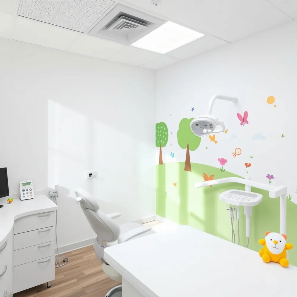
Patient comfort has markedly improved. Modern CMOS sensors are smaller, thinner, and more flexible, reducing gag reflexes and chair time. Lead‑free, child‑size aprons and thyroid collars are lightweight, making the experience less intimidating. Wireless handheld units remove cumbersome cables, creating a calmer environment that encourages cooperation from young patients.

Modern pediatric offices use digital intra‑oral sensors—often Silicon photodiode (CMOS) sensors are smaller and more flexible than CCD sensors—to capture high‑resolution images in a fraction of a second. These sensors reduce the radiation dose by 70‑90 % compared with traditional film, allowing clinicians to obtain clear pictures of early carious lesions while keeping exposure well below the levels of natural background radiation. Protective measures include Lead‑free pediatric‑size aprons and thyroid collars improve comfort and compliance. All imaging follows the ALARA (As Low As Reasonably Achievable) principle: the smallest field of view and lowest effective settings are selected, and an X‑ray is taken only when the diagnostic benefit outweighs any risk. Safety considerations for babies and toddlers therefore combine ultra‑low‑dose digital technology, child‑specific shielding, and strict adherence to pediatric guidelines that recommend radiographs only after primary molars begin to contact or when a specific clinical problem necessitates imaging.

Primary tooth eruption timeline – By 6‑12 months the first primary incisors appear; by 24 months all 20 baby teeth should be present.
What is the rule of 4 in pediatric dentistry? – The rule of 4 (or 7‑4 rule) links age in months to expected primary teeth. Subtract 4 from the child’s age in months to estimate tooth count (e.g., a 15‑month‑old should have ~11 teeth). By two years the full set of 20 is complete. This helps spot eruption delays early.
What is the rule of 7 in pediatric dentistry? – Children should receive their first comprehensive orthodontic evaluation around seven years of age to identify bite issues or crowding that may benefit from early intervention.
Why is early dental care important for children? – Establishing a dental home enables early detection of caries, proper fluoride and sealant use, and education on hygiene, preserving space for permanent teeth, supporting nutrition, speech, and reducing future pain, anxiety, and costly treatments.

Dental fillings are routinely placed in 3‑year‑old patients when a qualified pediatric dentist uses child‑friendly protocols. Modern tooth‑colored materials—composite resin and glass‑ionomer—are mercury‑free, bond well to primary teeth, and glass‑‑mer which release fluoride, which protects the tooth and surrounding enamel. A quick digital radiograph, which emits up to 90 % less radiation than film, may be taken to confirm the lesion while adhering to the ALARA principle. Pain is managed with the 3‑3‑3 rule (three 200 mg ibuprofen tablets every three hours for up to three days) and gentle local anesthesia; nitrous oxide or other light sedation options keep the child calm with discomfort. Fluoride varnish is often applied during the same visit to strengthen enamel and reduce future decay. These combined techniques allow safe, efficient restorative care that prevents pain, infection, and disruption of normal dental development.
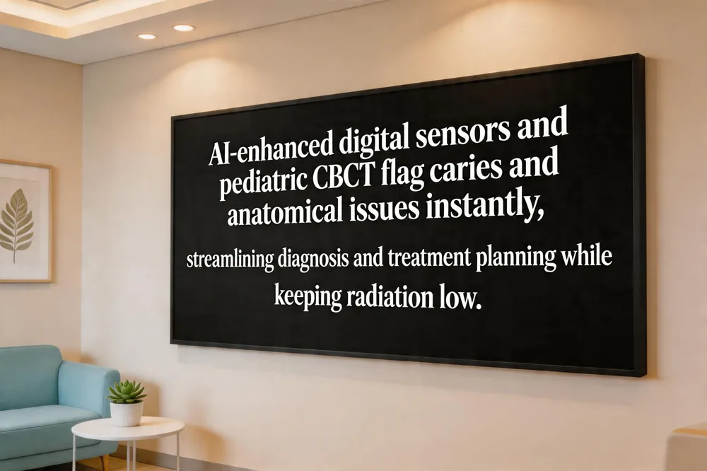
Modern pediatric dental imaging blends high‑resolution digital intra‑oral sensors, low‑dose cone‑beam computed tomography (CBCT), and AI algorithms. Digital sensors capture instant 2‑D images with up to 90 % less radiation than film, while pediatric‑specific CBCT provides 3‑D views at doses comparable to a few bitewings. Integrated AI flags potential pathology—caries, root resorption, bone loss—in seconds, offering color‑coded overlays and quantitative measurements that act as a reliable second opinion. AI‑enhanced CBCT can automatically segment teeth and assess spatial relationships, streamlining orthodontic and surgical planning. Tele‑radiology platforms securely share these enhanced images with specialists, reducing repeat scans and accelerating treatment decisions. Together, these advances lower radiation exposure, improve diagnostic accuracy, and increase workflow efficiency, delivering safer, faster care for children.

The dentist’s “rule of 2” reinforces everyday oral‑health habits: visit the dentist twice a year, brush twice daily, and brush for two minutes each time. Consistently following these simple steps supports healthy smiles and complements the high‑tech, child‑friendly environment created by VR distraction.
Future pediatric dental care will be driven by relentless imaging innovation that keeps radiation at the lowest possible levels while expanding diagnostic insight. At Gentle Dentistry of Staten Island, the latest low‑dose CBCT protocols, CMOS intra‑oral sensors, and wireless handheld units already deliver instant, high‑resolution images that children can tolerate with minimal gag reflex. Integrated AI tools flag early caries and developmental anomalies in real time, reducing repeat scans and accelerating treatment planning. Together with lead‑free aprons, thyroid collars, and strict ALARA monitoring, these advances ensure every child receives a safe, comfortable, and patient‑centered experience that supports lifelong oral health.