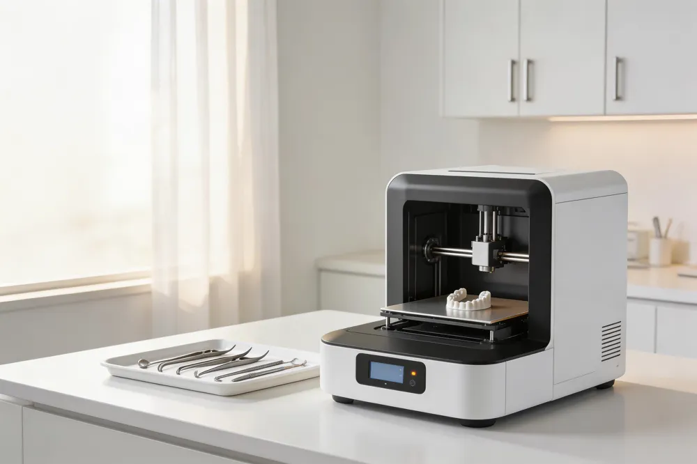
Even more knowledge

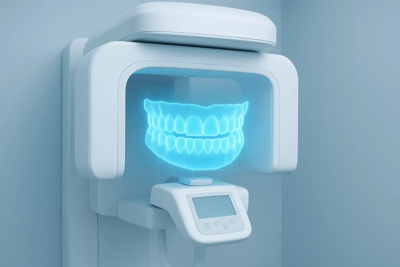
Cone Beam Computed Tomography (CBCT) is a specialized imaging technology that has revolutionized how dental professionals diagnose and plan treatment. Unlike traditional two-dimensional X-rays, a CBCT scan rotates a cone-shaped X-ray beam around your head to produce detailed three-dimensional images of your teeth, jawbone, nerves, and sinuses in a single, quick scan.
Traditional X-rays are excellent for routine checkups but have limitations. They can miss subtle issues hidden by overlapping teeth or provide an incomplete picture of complex anatomy. CBCT is specifically designed for situations where those standard images are not sufficient, offering a powerful, three-dimensional view that eliminates guesswork.
This advanced technology allows our practice to move beyond simple diagnosis into precise treatment simulation. With a clear, 360-degree understanding of your unique anatomy, we can develop highly personalized treatment plans. This leads to greater predictability, safety, and confidence for both the clinician and patient, especially for complex procedures.
The following article will delve into the specific advantages of CBCT imaging, from improved diagnostic accuracy for hidden infections to safer surgical planning for dental implants. We will also cover its responsible application across various specialties, ensuring this powerful tool is used wisely to directly benefit your oral health.
Cone Beam Computed Tomography (CBCT) is an advanced dental imaging technology. Unlike standard X-rays, it uses a rotating, cone-shaped X-ray beam to capture a large series of images from multiple angles. These images are then digitally reconstructed into a highly detailed, three-dimensional model of your teeth, jawbones, nerve canals, and sinuses.
A CBCT scan is a quick and painless procedure. For a full mouth scan, you remain still for just 20 to 40 seconds while the imaging device rotates around your head. When focusing on a specific region, the scan can take less than 10 seconds. There is no special preparation needed, and the process is non-invasive.
Traditional dental X-rays, like bitewings or panoramic images, produce a flat, two-dimensional picture. This can cause structures to overlap or hide complex anatomy. CBCT eliminates this problem of superimposition. It provides a clear, volumetric 3D view, allowing dentists to examine bone width, measure nerve proximity, and view structures from any angle.
Historically, dentists had to refer patients to hospitals for medical CT scans when 3D imaging was needed. CBCT technology was developed in the late 1990s to address this. The machines are more compact, affordable, and produce images optimized for the jaw and face. This evolution made powerful 3D imaging a practical, in-office tool for dental specialists.
The primary benefit of CBCT is its ability to provide a comprehensive, detailed view of hard tissues in a single scan. It excels at imaging bone density, tooth roots, and critical anatomical landmarks. This clarity is crucial for complex procedures where precision is paramount, such as planning dental implants or diagnosing hidden dental fractures.
Essential Facts About CBCT Technology
| Aspect | Key Detail | Why It Matters |
|---|---|---|
| Scanning Technology | Uses a rotating cone-shaped X-ray beam and a 2D detector. | This design captures a full volume of data in a single rotation, creating the 3D model. |
| Radiation Exposure | Delivers a lower radiation dose than a medical CT but more than a standard dental X-ray. | Dentists follow the "ALARA" principle—using the lowest dose reasonably achievable for diagnosis. |
| Primary Applications | Crucial for implant planning, evaluating impacted teeth, complex root canal anatomy, jaw pathology, and trauma assessment. | It provides the detailed 3D information necessary for safe and predictable treatment planning in these areas. |

The fundamental limitation of traditional two-dimensional dental X-rays is superimposition. Overlapping anatomical structures can hide critical details like cavities between teeth, fractures, or the exact location of an impacted tooth.
Cone Beam Computed Tomography (CBCT) solves this problem by providing a three-dimensional, volumetric image. This allows dentists to visualize anatomy in three planes—horizontal, vertical, and axial—providing crucial depth and density information that is impossible to obtain from a flat picture.
Research consistently demonstrates CBCT's superiority over conventional periapical radiography for several key diagnostic tasks.
A review of studies found CBCT to be clearly superior for detecting vertical root fractures and periapical lesions. For bone defects, such as those associated with periodontal disease, CBCT also provides more accurate assessments than 2D images.
Specifically, CBCT offers a clear advantage in visualizing the buccolingual (cheek-to-tongue) width of bone, a critical measurement for procedures like dental implant placement with CBCT. This three-dimensional data eliminates the distortion and magnification errors common in panoramic or periapical X-rays.
The enhanced detail from CBCT directly influences treatment planning, especially in complex cases. Systematic reviews show that CBCT often changes the diagnosis and treatment plan in endodontics, particularly for dental trauma, external root resorption, and complex root canal anatomy.
Studies have documented that clinicians using CBCT are more definitive in their treatment choices. When presented with a trauma case, professionals were more likely to propose a clinical intervention, such as starting conventional root canal treatment, after reviewing a CBCT scan compared to a standard periapical X-ray.
Perhaps one of the most significant advantages is the elimination of guesswork. CBCT provides a precise, measurable map of the oral landscape.
For implant surgery, surgeons can see the exact proximity of the inferior alveolar nerve canal or the maxillary sinus floor to within a fraction of a millimeter. This allows for the selection of the perfect implant length and angulation, drastically reducing the risk of nerve injury or sinus perforation.
Similarly, for impacted wisdom teeth, CBCT can clarify the tooth's relationship to the nerve canal in three dimensions, although current guidelines recommend its use only when 2D imaging leaves critical questions unanswered.
Despite its advanced capabilities, CBCT is not intended to replace conventional dental X-rays for all purposes. Traditional bitewing and periapical radiographs remain the first-line, low-dose imaging choice for routine dental care.
They are perfectly suited and highly effective for detecting cavities between teeth, monitoring bone levels in gum disease, and checking for infections during regular check-ups. The guiding principle, known as ALARA (As Low As Reasonably Achievable), dictates using the lowest radiation dose necessary to obtain the required diagnostic information.
Therefore, for simple diagnostics and routine monitoring, the minimal radiation of a traditional X-ray is appropriate. CBCT is reserved for complex diagnostic challenges and precise pre-surgical planning where its three-dimensional insight provides a decisive clinical benefit.
| Diagnostic Scenario | Traditional 2D X-Rays | Cone Beam CT (CBCT) | Primary Advantage of CBCT |
|---|---|---|---|
| Cavity Detection Between Teeth | Primary method; sufficient detail | Not recommended for routine use; lower resolution | N/A - 2D is preferred |
| Vertical Root Fracture | Often missed due to superimposition | High sensitivity and specificity | Clear 3D visualization eliminates overlap |
| Dental Implant Planning | Limited; no buccolingual bone width data | Gold standard; allows precise 3D bone mapping | Enables virtual surgery and risk avoidance |
| Complex Root Canal Anatomy | Can miss extra canals or unusual shapes | Reveals true 3D morphology of root canal system | Leads to more complete treatment |
| Impacted Tooth Assessment | Shows basic position and angulation | Shows precise 3D relationship to nerves and adjacent teeth | Provides critical spatial data for safe extraction |
| Periodontal Bone Defects | Shows general bone level changes | Accurately measures defect depth, morphology, and furcation involvement | Enables more targeted and predictable treatment |

At Gentle Dentistry of Staten Island, Cone Beam Computed Tomography (CBCT) is reserved for specific, complex cases where traditional 2D imaging is insufficient. Its ability to provide detailed three-dimensional views makes it invaluable for precise treatment planning across several dental specialties.
CBCT is the imaging modality of choice for planning dental implants. It allows our specialists to accurately measure bone height, width, and density. This technology helps identify vital structures like nerves and sinuses, enabling the creation of precise surgical guides. This meticulous planning minimizes risks and ensures optimal implant placement.
For intricate root canal treatments, a small-field CBCT scan provides clarity when periapical X-rays are ambiguous. It is particularly useful for visualizing complex root canal anatomy, detecting additional canals, and assessing root resorption or fractures. This detailed information directly influences diagnosis and can change the treatment plan, leading to more predictable outcomes.
In orthodontics, CBCT is ideal for evaluating the position of unerupted teeth, such as canines, and assessing their relationship to nerves. For impacted third molars (wisdom teeth), CBCT is not routine but is used when 2D images cannot answer specific questions about a tooth's position relative to critical anatomy, aiding in safer surgical planning.
CBCT excels at evaluating cysts, benign tumors, and fractures of the jaws. It provides a clear view of the lesion's extent and its proximity to vital structures. For procedures like sinus lift bone grafting, CBCT is recommended preoperatively to assess membrane thickness, sinus anatomy, and residual bone height, which is crucial for success.
CBCT is highly effective for detecting bony changes in the jaw joint, such as those caused by osteoarthritis. However, for evaluating soft tissue problems like disc displacement, magnetic resonance imaging (MRI) remains the preferred method, as CBCT provides limited soft tissue detail.
Studies show CBCT significantly improves diagnosis and treatment planning for dental trauma. It is superior to conventional X-rays for detecting root fractures and evaluating the extent of alveolar bone injuries. This leads to more definitive treatment decisions and increased clinician confidence in managing traumatic injuries.
| Clinical Application | Primary Diagnostic Use | Key Benefit over 2D Imaging |
|---|---|---|
| Implant Planning | Assessing bone volume & nerve proximity | Precise 3D measurements for guided surgery |
| Complex Endodontics | Visualizing root anatomy & fractures | Reveals extra canals and hidden pathology |
| Oral Surgery/Pathology | Evaluating cysts, tumors, & jaw fractures | Shows true lesion extent in three dimensions |
| Dental Trauma | Diagnosing root & alveolar bone fractures | Higher detection rate for fine fractures |
| Orthodontics | Locating impacted/unerupted teeth | Spatial relationship to nerves is clear |
| TMJ Disorders | Assessing condylar bone changes | Excellent detail for arthritic changes |

The effective radiation dose from a dental CBCT scan is higher than that from a single conventional dental X-ray, such as a bitewing or periapical radiograph. However, its dose is significantly lower—up to 98.5% less—than that of a conventional medical CT scan. For context, CBCT effective doses can range from approximately 11 to 477 microsieverts (μSv). This is comparable to just a few days of exposure to natural background radiation.
Importantly, the radiation used in a CBCT scan is external; no radiation remains in your body after the examination. The procedure itself is quick, painless, and non-invasive, with a full-mouth scan typically taking 20 to 40 seconds.
To ensure patient safety, dentists follow the ALARA principle—"As Low As Reasonably Achievable." This means radiation exposure is minimized by using the lowest possible dose to obtain the necessary diagnostic information. Consequently, CBCT is not used for routine screening. Its use must be justified by a specific clinical need where the benefits of the 3D information outweigh the small radiation risk.
Key to the ALARA principle is selecting the smallest necessary Field of View (FOV) for the specific diagnostic task. A smaller FOV targets only the area of interest, reducing the patient's overall radiation exposure. For most basic dental diagnostic needs, such as detecting cavities or monitoring gum health, traditional 2D imaging (like bitewings and panoramic X-rays) remains the first and most appropriate choice due to their lower radiation dose.
| Imaging Modality | Typical Effective Dose Range | Contextual Comparison | Primary Justification for Use |
|---|---|---|---|
| Single Digital Dental X-ray | ~5-6 μSv | Equivalent to ~1 day of background radiation | First-line imaging for basic diagnosis (cavities, bone levels) |
| Dental CBCT Scan | ~11-477 μSv | Equivalent to a few days of background radiation; up to 98.5% less than medical CT | Complex cases requiring 3D assessment (implants, surgery, trauma) |
| Medical CT Scan (Head/Neck) | ~2000 μSv | Significantly higher radiation exposure | Broad medical imaging needs beyond the dental scope |
The dental community emphasizes extra caution when imaging younger patients, who are more sensitive to radiation. The "Image Gently" campaign is dedicated to educating professionals about optimizing radiation techniques specifically for pediatric patients, reinforcing the commitment to the ALARA principle across all age groups.
A primary limitation of Cone Beam Computed Tomography (CBCT) is its relatively poor soft tissue contrast. While CBCT provides exceptional detail of bony structures, it cannot clearly delineate muscles, lymph nodes, or glands. This makes it unsuitable for evaluating suspected malignancies or pathologies primarily involving soft tissues. For detailed soft tissue analysis, as in cases of temporomandibular joint (TMJ) disc disorders, Magnetic Resonance Imaging (MRI) is the preferred modality.
CBCT is not intended for routine dental screening. Professional guidelines strongly recommend against its use for routine caries detection or for the initial assessment of periodontal disease. For these purposes, conventional intraoral radiographs like bitewings remain the standard. Similarly, CBCT is not routinely recommended for assessing impacted mandibular third molars (wisdom teeth), as current evidence shows it does not reduce the risk of nerve injury compared to panoramic radiographs. It should only be considered if a specific clinical question, such as the precise relationship of a root to the mandibular nerve canal, cannot be answered by two-dimensional imaging.
Image quality in CBCT can be degraded by various artifacts. Metallic restorations like amalgam fillings or crowns can cause significant streaking or distortion, potentially obscuring important anatomical details. Patient movement during the scan, which typically lasts between 5 to 40 seconds, can also result in motion blur. Furthermore, the technology has inherent limitations in contrast resolution compared to conventional medical CT scanners. This means subtle density differences in bone or adjacent structures may not be as clearly visualized.
CBCT involves a higher effective radiation dose than conventional dental X-rays, though it is lower than traditional medical CT. The dose varies significantly based on the field of view (FOV) and scanner settings. Therefore, a fundamental principle for its use is justification. Authorities like the American Association of Endodontists (AAE) and the American Academy of Oral and Maxillofacial Radiology (AAOMR) stipulate that CBCT should only be used when the diagnostic benefit is likely to change the patient's treatment plan and outweighs the radiation risk. Following the ALARA principle (As Low As Reasonably Achievable), clinicians should always select the smallest possible FOV to accomplish the specific diagnostic task.
| Consideration | Primary Limitation | Clinical Implication |
|---|---|---|
| Soft Tissue Detail | Poor contrast resolution for muscles, glands, and tumors. | Not suitable for evaluating suspected cancer or TMJ disc issues; MRI is superior. |
| Routine Diagnostics | Higher radiation dose than conventional X-rays. | Not recommended for routine caries detection or initial periodontal evaluation. |
| Image Quality | Susceptible to metal artifacts from fillings and patient motion. | May compromise diagnosis in heavily restored dentition; patient cooperation is critical. |
| Clinical Justification | Benefits must demonstrably outweigh the risks per professional guidelines. | Should be reserved for complex cases in endodontics, implantology, and trauma where 2D imaging is insufficient. |
Dental insurance claims for Cone Beam CT (CBCT) scans are processed using Current Dental Terminology (CDT) codes. The primary code for a comprehensive full-mouth scan is D0367, which applies to imaging both jaws, often including the cranium. For a scan focused on just the upper or lower jaw, providers use D0365. A more targeted, limited field-of-view scan—ideal for evaluating a single problematic area—is billed under D0364. In advanced workflows, dentists may fuse a CBCT scan with an optical surface scan for precise virtual planning; this specialized service is coded as D0395.
Dental insurance coverage for CBCT scans is not universal. It is highly dependent on the individual policy and, crucially, on whether the scan is deemed medically necessary for diagnosis or treatment planning. Major insurers, including Delta Dental, have recently updated their policies to include CBCT coverage, reflecting its growing integration into standard care. However, approval often hinges on the dentist providing a detailed justification to the insurance company.
Insurance is most likely to cover a CBCT scan when it is essential for complex, non-routine procedures. Typical scenarios include preoperative planning for dental implant placement to assess bone quality and vital structure proximity. It also includes evaluating complex root canal anatomy, diagnosing dental trauma like root fractures, or investigating jaw pathologies such as cysts or tumors. These situations demonstrate a clear diagnostic need that cannot be fully met with conventional 2D X-rays.
CBCT scans are less likely to be covered for routine, simple procedures. Standard diagnostic needs, such as planning for a single crown or a conventional bridge, are usually adequately assessed with traditional intraoral X-rays. Insurance companies typically view CBCT in these contexts as providing incremental, rather than essential, information and may deny the claim.
The out-of-pocket cost for a CBCT scan should be viewed as a strategic investment in your care. For complex treatments, the detailed three-dimensional visualization translates into superior precision. It allows for meticulous surgical planning, which significantly reduces risks like nerve injury or sinus perforation. This enhanced planning leads to more predictable outcomes, shorter procedure times, and potentially fewer complications. Ultimately, the upfront cost supports a higher standard of safety and long-term success for major dental work.
| CDT Code | Clinical Application | Typical Scan Scope |
|---|---|---|
| D0367 | Comprehensive Treatment Planning | Both jaws, full arch |
| D0365 | Single-Jaw Evaluation | Upper or lower arch only |
| D0364 | Focused Problem Area | Few teeth, limited view |
| D0395 | Advanced Fused Imaging | CBCT + intraoral scan data |
It provides clinicians with a level of anatomical detail unattainable with traditional 2D radiography. The ability to visualize structures in three dimensions—to see around corners, measure bone width precisely, or detect fine root fractures—has transformed treatment planning for procedures ranging from complex root canals to the precise placement of dental implants.
Professional guidelines consistently emphasize that CBCT should not replace conventional radiography for routine examinations. Its use is justified when a diagnosis cannot be made with standard X-rays and when the clinical benefits demonstrably outweigh the slightly higher radiation exposure. This adherence to the ALARA (As Low As Reasonably Achievable) principle is a cornerstone of responsible radiology.
Clinics that integrate CBCT, like Gentle Dentistry, do so to elevate patient care in specific scenarios. The technology enhances diagnostic confidence in ambiguous cases, improves procedural safety by mapping out vital structures, and enables the creation of highly personalized treatment plans. This leads to more predictable outcomes, reduced risk of complications, and ultimately, better long-term oral health.
Ultimately, CBCT is a testament to dentistry's progress. It is a powerful tool wielded with wisdom, reserved for situations where its detailed insight can directly improve patient outcomes. Its strategic use represents a commitment to combining clinical expertise with cutting-edge technology to provide the most informed, effective, and safe care possible.