Even more knowledge


Periodontal pocket depth is the distance measured from the gingival margin to the base of the sulcus with a calibrated probe; healthy sites measure 1–3 mm, while depths of ≥4 mm indicate disease and deeper pockets (≥5‑6 mm) signal moderate to severe periodontitis. Persistent deep pockets trap plaque and subgingival biofilm, perpetuating inflammation, bone loss, and tooth mobility; they also increase systemic risk, linking gum disease to cardiovascular disease, diabetes complications, and adverse pregnancy outcomes. The primary therapeutic goal is to shrink these pockets to a healthy ≤3 mm without bleeding on probing, thereby halting bacterial colonization, allowing re‑attachment of gingival tissue, and preserving bone support. Achieving this requires a step‑wise approach: meticulous non‑surgical debridement (scaling and root planing), adjunctive antimicrobial or host‑modulation agents, and, when necessary, surgical pocket‑reduction or regenerative procedures. Long‑term stability depends on diligent home oral hygiene, regular supportive periodontal therapy, and management of systemic risk factors such as smoking and diabetes.

Scaling and root planing (SRP) remains the first‑line non‑surgical treatment for periodontitis. By mechanically removing subgingival plaque and calculus and smoothing root surfaces, SRP reduces probing pocket depths 1–2 mm in most patients and promotes gingival re‑attachment. Adjunctive antimicrobial agents—such as locally delivered antibiotics, chlorhexidine chips, or systemic amoxicillin/metronidazole—can enhance this effect, especially in deeper sites. The Laser‑Assisted New Attachment Procedure (LANAP) uses a 1064‑nm Nd:YAG laser to selectively eliminate diseased epithelium and biofilm while preserving healthy tissue. Clinical studies report LANAP achieving pocket‑depth reductions of 1–3 mm after a single session, often surpassing SRP alone, and may stimulate bone regeneration.
Is LANAP better than scaling and root planing? Yes. Evidence shows LANAP provides greater improvements in attachment levels, pocket reduction, inflammation control, and bone fill compared with SRP alone, while being less invasive and offering quicker recovery. SRP is still essential for mild to moderate disease or as a prerequisite before LANAP.
Can 3 mm gum pockets be reversed? A 3 mm pocket is already within the healthy range (1–3 mm) and can be maintained or slightly reduced through diligent oral hygiene and regular professional cleanings. If inflammation has temporarily deepened the sulcus, SRP often restores it to normal depth, and ongoing maintenance prevents recurrence.
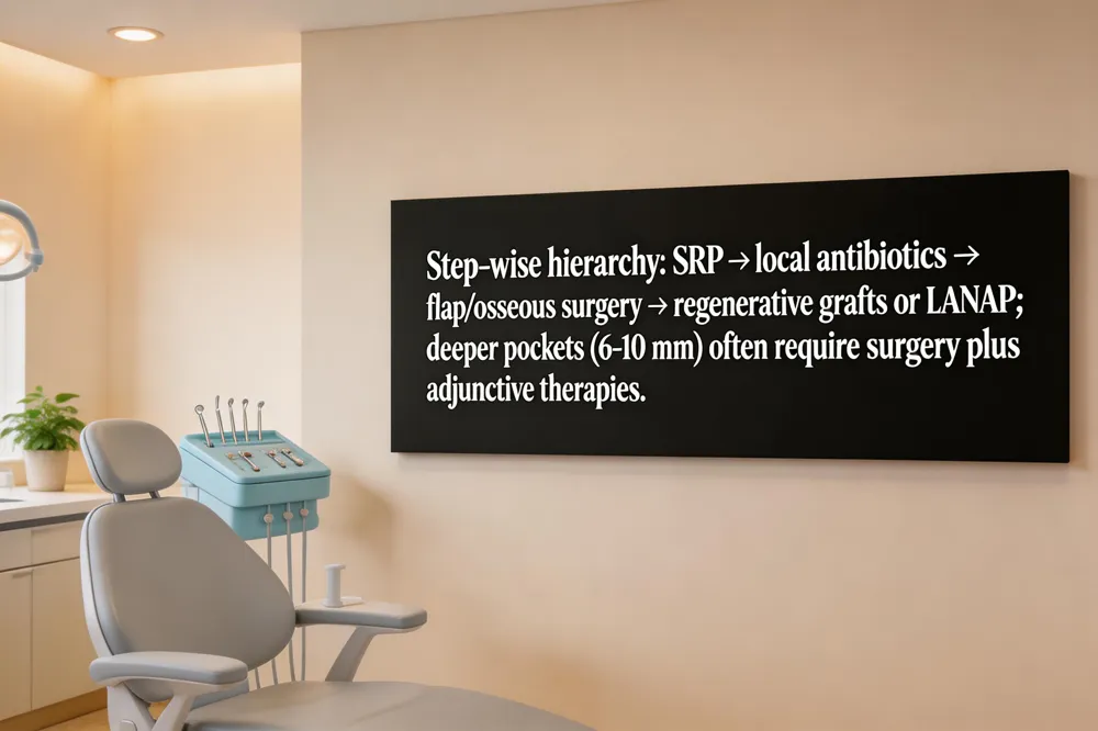
Step‑wise treatment hierarchy – Begin with thorough oral‑hygiene instruction and scaling & root planing (SRP). If probing depths remain ≥4 mm, add locally delivered antibiotics or antimicrobial rinses; reassess at 4–6 weeks.
Surgical flap and osseous techniques – For persistent pockets ≥5 mm, perform flap (pocket‑reduction) surgery, often combined with osseous recontouring to eliminate bony irregularities and enable primary closure.
Regenerative approaches – When bone loss is evident, place bone grafts or use guided tissue regeneration (GTR) membranes; platelet‑rich fibrin (PRF) can enhance soft‑tissue healing and attachment.
Adjunctive antibiotics and laser therapy – Local antibiotics (e.g., minocycline microspheres) or short‑course systemic agents reduce residual microbes; laser‑assisted new‑attachment procedures (LANAP) or Nd:YAG debridement may further shrink pockets.
10 mm pockets – SRP + local antibiotics, then flap‑osseous surgery with GTR or LANAP; grafts often required.
8 mm pockets – SRP, flap surgery with bone grafts or GTR; adjunctive antibiotics/laser.
6 mm pockets – SRP can close many; if not, minimally invasive flap or laser therapy; maintenance critical.
5 mm pockets – SRP plus meticulous home care; localized antibiotics; surgery if needed.
4 mm pockets – Professional SRP and maintenance; home care alone rarely closes pocket; adjunctive antimicrobials help.

Impact of tooth type, root number, and furcation involvement
Molars and multirooted teeth exhibit smaller probing‑pocket‑depth (PPD) reductions than single‑rooted incisors or premolars. Presence of furcation involvement (grade 2/3) further limits depth gain, because the complex anatomy hinders thorough debridement.
Role of restorations, mobility, and plaque index
Teeth with crowns or large fillings start with higher baseline PPD and achieve less closure (≈1.0 mm vs ≈1.4 mm for unrestored teeth). Mobility does not change the absolute reduction but reduces the odds of complete closure. Plaque‑positive sites lose on average 0.2 mm more depth than plaque‑negative sites, underscoring the importance of oral‑hygiene adherence.
Multilevel logistic regression findings
A multilevel model of 16,825 teeth identified independent predictors of pocket closure: baseline PPD 4‑5 mm (OR 4.1), single‑rooted anatomy (OR 1.9), absence of restoration (OR 1.4), furcation grade 0/1 (OR 1.3), lack of mobility (OR 1.2), and plaque‑negative status (OR 1.3).
Probing pocket depth reduction after non‑surgical periodontal therapy: tooth‑related factors
Non‑surgical therapy lowers overall PPD by ~1.2 ± 1.5 mm, with deeper pockets (>6 mm) showing the greatest absolute drop but often remaining deep. Tooth‑specific factors—type, root count, furcation, vitality, mobility, and restorations—significantly influence closure rates. Recognizing these variables before treatment helps clinicians anticipate which sites may need re‑instrumentation or surgery.
Periodontal treatment before and after
Patients present with swollen, bleeding gums and bad breath. During treatment, scaling and root planing (often complemented by laser or adjunctive antibiotics) removes subgingival biofilm, smoothing roots and preparing tissues for reattachment. Post‑treatment gums become firmer, pink, and less prone to bleeding; patients follow gentle brushing, a short soft‑food diet, and regular maintenance visits to preserve the achieved depth reductions.
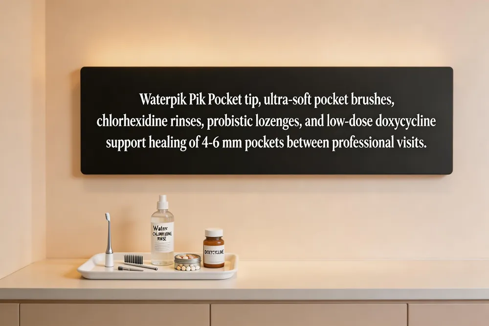
Effective pocket reduction begins with a professional deep‑cleaning (scaling and root planing) that eliminates subgingival plaque and tartar. At home, the Waterpik Pik Pocket tip can irrigate pockets up to 6 mm deep, flushing out debris and bacteria when used twice daily with a low‑pressure setting and an antimicrobial rinse (chlorhexidine or salt‑water). A specialized gum‑pocket brush—tiny, ultra‑soft bristles—reaches the narrow space between gum and tooth, disrupting biofilm that regular brushes miss; use it once a day after brushing. Antimicrobial rinses such as chlorhexidine (diluted hydrogen peroxide, or natural sage/tea‑tree oil solutions) control bacterial load, while probiotic lozenges (e.g., Lactobacillus reutri) and host‑modulation agents like sub‑antimicrobial dose doxycycline modestly reduce inflammation and matrix metalloproteinase activity. Maintaining this regimen, combined with regular flossing or interdental brushes, supports the healing of 4 mm pockets and can reverse moderate disease when professional care is promptly followed up with quarterly maintenance visits.

The newest approach blends regenerative medicine with minimally invasive tools. Stem‑cell therapy combined with 3‑D‑printed scaffolds is used to regrow lost bone and gingival tissue, while laser‑assisted protocols such as LANAP selectively remove diseased epithelium with minimal bleeding. Antimicrobial photodynamic therapy (aPDT) delivers a light‑activated photosensitizer that destroys pathogens without antibiotics. AI‑driven cone‑beam CT and salivary biomarker panels (IL‑1β, MMP‑8) pinpoint active sites early, allowing clinicians to tailor these advanced modalities for each patient.
In the United States, stem‑cell gum regeneration typically starts around $4,000 for a single injection and can range $15,000–$30,000 for a full protocol, compared with $2,000–$8,000 for LANAP. Costs reflect cell source, laboratory processing, number of sessions, and geographic location.
LANAP was introduced in 1994, making it roughly 30 years old. Over three decades it has evolved with improved Nd:YAG lasers and refined protocols, becoming a standard flap‑less option for periodontitis.
Clinical data show LANAP achieves greater probing‑depth reductions than conventional non‑surgical therapy, often decreasing pockets by 1–3 mm and reducing Porphyromonas gingivalis levels.
Emerging senolytics aim to clear senescent cells in inflamed periodontal tissues, potentially enhancing healing and supporting the regenerative effects of stem‑cell scaffolds. Ongoing trials in 2024 are evaluating their adjunctive benefit to pocket‑depth outcomes.
Effective management of periodontal pockets follows a step‑wise pathway: early detection, thorough oral‑hygiene instruction, and non‑surgical debridement (scaling and root planing) that typically reduces probing depths by 1–2 mm. When pockets remain ≥4 mm, adjunctive measures—locally delivered antibiotics, antimicrobial rinses, or laser‑assisted therapy—can enhance outcomes. Persistent deep sites (≥6 mm) often require minimally invasive flap surgery, bone‑shaping, or regenerative grafts to achieve further depth reduction and promote re‑attachment. Gentle Dentistry of Staten Island tailors each phase to the patient’s specific tooth‑related factors (tooth type, root number, furcation involvement, restoration status, plaque control, and mobility), as demonstrated by recent multilevel regression studies. This personalized approach improves prediction of sites that may need re‑instrumentation or surgery and maximizes long‑term stability. If you notice bleeding, persistent bad breath, or deeper pockets, schedule a professional evaluation at Gentle Dentistry of Staten Island to develop a customized treatment plan and protect your gum health.