Even more knowledge

In the United States, about 21 % of adults aged 20‑64 have untreated cavities, a chronic condition that can progress to pain, infection, and tooth loss if not caught early. Detecting decay at the enamel or dentin stage enables minimally invasive interventions—such as fluoride varnish, sealants, remineralization therapies, or micro‑restorations—that preserve natural tooth structure and often avoid the need for drilling or crowns. This approach dovetails with Gentle Dentistry of Staten Island’s patient‑centered philosophy: early, painless diagnosis, transparent visual communication (heat‑map overlays on radiographs), and treatment plans that prioritize comfort and preservation. By integrating AI‑driven radiographic analysis, the practice can identify lesions that are invisible to the naked eye, delivering timely, gentle care that aligns with its commitment to gentle, preventive dentistry.

Deep‑learning convolutional neural networks (CNNs) are the engine behind modern AI dental radiography. Trained on millions of expertly annotated bitewing, periapical and panoramic images, these networks learn to recognize subtle pixel‑level patterns of enamel demineralization, dentin involvement, bone loss and sinus proximity. At the point of capture the software first standardises the image, then runs a rapid object‑detection model—often a YOLO‑derived architecture—to flag suspicious areas. The output is presented as real‑time heat‑map overlays or colour‑coded bounding boxes that highlight the exact location and severity of a lesion, sometimes with quantitative risk scores or millimetre measurements.
Workflow integration is seamless: the AI engine runs in the background as the radiograph is uploaded, automatically attaching visual annotations to the patient record and suggesting diagnostic codes. Clinicians review the overlay, confirm findings, and incorporate them into treatment planning. This decision‑support reduces intra‑examiner variability, shortens interpretation time by up to 30%, and provides a clear visual aid for patient education—key for a gentle, patient‑centered practice like Gentle Dentistry of Staten Island. The result is faster, more consistent diagnoses that enable minimally invasive interventions and improve overall oral‑health outcomes.
Artificial intelligence in dental radiology pdf – The PDF titled “Artificial intelligence in dental radiology: a review” (Mar 2025, Annals of Medicine and Surgery 87(4)) provides a comprehensive overview of AI techniques, accuracy gains, and workflow integration.
AI in dental diagnosis – AI analyzes radiographs and intra‑oral scans with speed and precision, detecting early carious lesions, periodontal disease, and periapical pathology, supporting personalized, patient‑centered care.
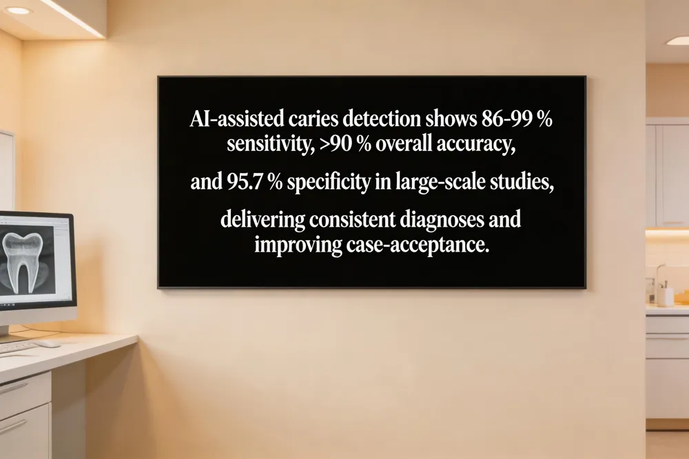
Can AI detect tooth decay? – Yes; studies show sensitivity 86‑99 % and overall accuracy >90 %, enabling minimally invasive interventions.
AI caries detection software – Real‑time overlays from FDA‑cleared AI (e.g., Overjet, Dentrix Detect AI) improve diagnostic consistency and patient communication, streamlining case acceptance.
Diagnostic accuracy of AI‑assisted caries detection – In a clinical evaluation of 4,361 teeth, AI achieved 93.4 % overall accuracy, 81.3 % sensitivity, and 95.7 % specificity, confirming its reliability as a decision‑support tool while highlighting areas for further improvement.
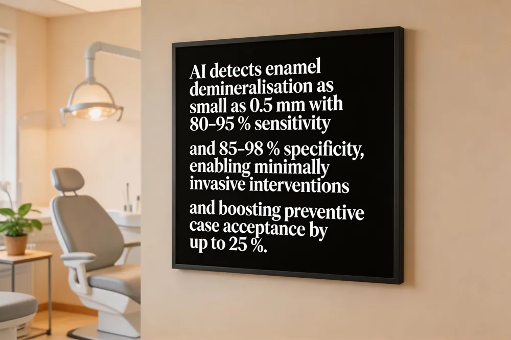
Artificial intelligence (AI) is rapidly transforming early childhood caries (ECC) detection and management. Deep‑learning convolutional neural networks trained on millions of annotated bitewing radiographs and intra‑oral photographs can spot enamel demineralization as small as 0.5 mm, delivering sensitivities of 80‑95 % and specificities of 85‑98 %—often surpassing manual interpretation. Real‑time heat‑map overlays highlight suspicious surfaces, allowing clinicians to verify findings instantly and reducing intra‑examiner variability. Early AI‑driven identification enables minimally invasive interventions such as fluoride varnish, sealants, or remineralization therapies that preserve tooth structure and lower long‑term costs. Visual AI overlays also improve patient education and shared decision‑making, boosting case acceptance by up to 25 % in practices that employ them. Regulatory clearance (FDA, ADA/ANSI standards) confirms safety and efficacy, while integration with practice‑management software streamlines documentation and billing. By adopting AI tools, pediatric dental practices can diagnose ECC earlier, offer gentler, patient‑centered care, and ultimately improve oral‑health outcomes for children.
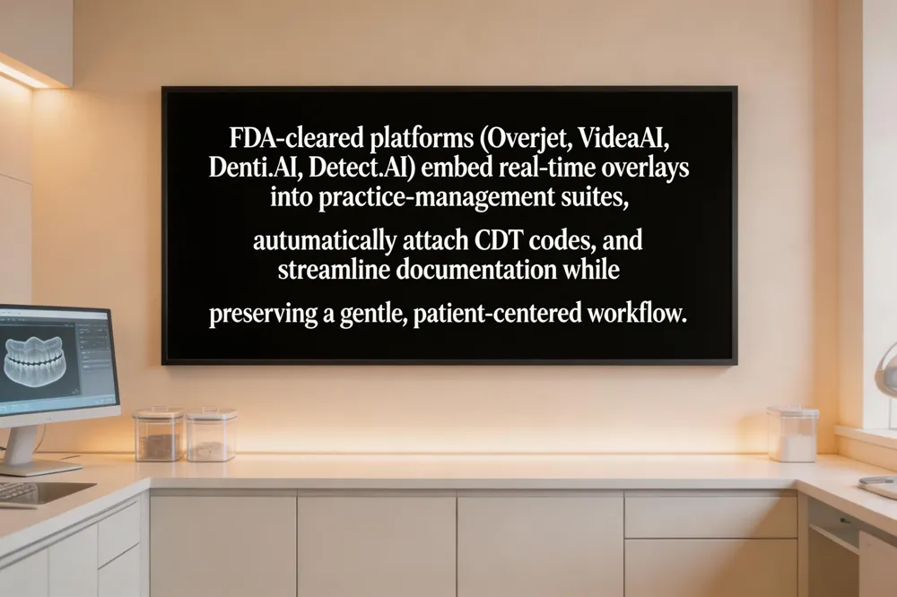
AI platforms cleared for dental use such as Overjet, VideaAI, Denti.AI, and Detect AI have FDA clearance and are validated across diverse radiographic equipment, ensuring safety and efficacy in clinical settings. These tools embed directly into practice‑management suites (Dentrix, Curve Dental, Ascend) and automatically attach CDT codes, streamlining documentation and insurance claims while preserving a gentle, patient‑centered workflow.
Seamless integration is achieved through real‑time overlays (heat maps, colour‑coded boxes) that appear on bitewing, periapical or panoramic images as they are captured, reducing interpretation time by up to 30 % and allowing clinicians to focus on communication and preventive counseling. The visual insights are patient‑friendly, helping hygienists and dentists explain early demineralisation, recommend minimally invasive treatments (fluoride varnish, sealants, Curodont), and improve case acceptance.
AI dental software download – The free AI:Dental app (Android, Google Play) offers annotated X‑ray libraries, quizzes, and on‑device AI analysis for education and skill‑building.
X‑ray analysis online free – Web‑based DICOM viewers like Medicai or CT Read provide instant AI‑generated interpretations for personal use.
AI caries app – Smartphone tools (e.g., SMARTeeth) analyse photos to flag early lesions, supporting parental monitoring and early preventive care.
AI radiology software – Integrated AI platforms triage urgent findings, generate preliminary reports, and reduce radiologist workload while maintaining regulatory compliance.
AI X‑ray app – Cloud‑based apps (IA X‑rays, qXR) deliver rapid diagnostics, secure storage, and multilingual reports, enhancing efficiency in any dental practice.
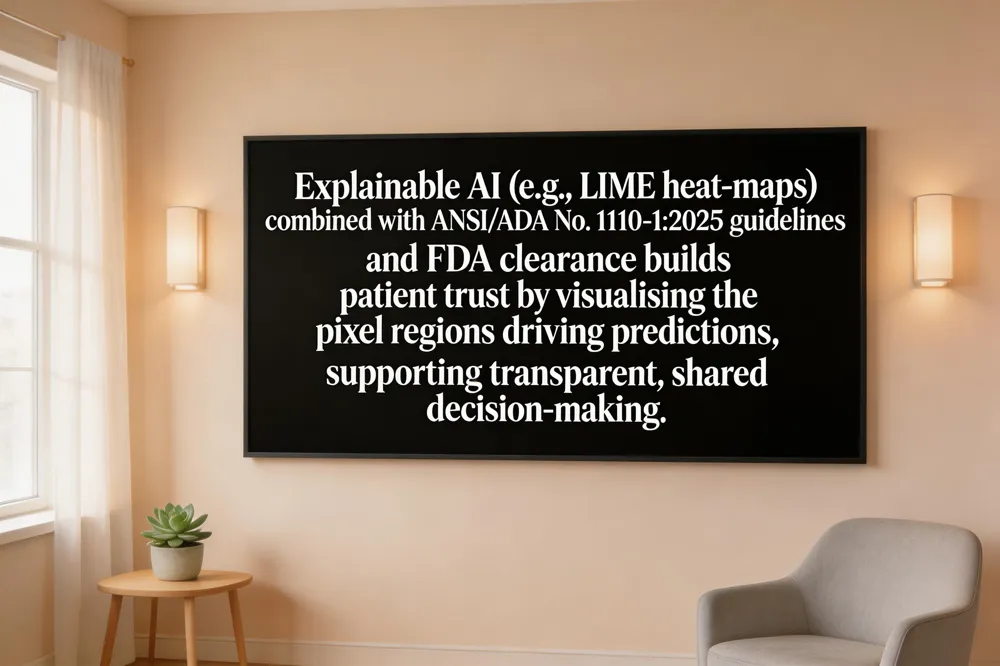
Explainable AI (XAI) is becoming essential for building patient trust in dental diagnostics. Deep‑learning models such as ResNet‑27 or YOLOv8 that detect carious lesions now embed XAI techniques (e.g., LIME heat‑maps) that visually highlight the exact pixel regions driving a positive prediction. This transparency lets clinicians show patients why a tiny enamel demineralization was flagged, turning a black‑box result into an educational conversation and fostering confidence in early, minimally invasive interventions.
Regulatory frameworks support safe adoption. The U.S. FDA has cleared AI‑based imaging tools such as Denti.AI, OverjetAI, and VideaAI, confirming their safety and efficacy. In parallel, the ANSI/ADA Standard No. 1110‑1:2025 provides uniform guidance for image annotation, data collection, and performance reporting, ensuring AI systems are fair, reproducible, and interoperable across digital radiography platforms used at Gentle Dentistry of Staten Island.
To translate these advances into everyday practice, comprehensive educational resources and staff training are required. Workshops on interpreting AI overlays, understanding sensitivity/specificity (often 85‑95% sensitivity, 90‑98% specificity for bite‑wing caries), and navigating practice‑management integration (e.g., Dentrix, Curve Dental) empower dentists, hygienists, and assistants to use AI as a decision‑support tool rather than a replacement. Ongoing education also includes reviewing ANSI/ADA guidelines and FDA labeling to maintain compliance and uphold the practice’s patient‑centered, gentle care philosophy.
AI algorithms, especially deep‑learning CNNs, now match or exceed the diagnostic performance of seasoned radiologists. Sensitivity for enamel and dentin caries on bitewing images regularly reaches 80‑95 % with specificity 85‑98 %, cutting false‑negative rates by up to 30 % and slashing interpretation time by roughly one‑third. By delivering heat‑map overlays and quantitative risk scores in real time, AI turns a static radiograph into an interactive guide that patients can see instantly. This visual dialogue supports Gentle Dentistry’s patient‑centered philosophy: clinicians can explain the exact location and severity of decay, propose minimally invasive preventive options, and involve the patient in shared decision‑making. The result is a gentler, more transparent experience that builds trust and improves oral‑health outcomes for both clinicians and families daily.