Even more knowledge

Dental radiography has undergone a fundamental transformation since the late 1980s. The transition from traditional screen-film systems to digital imaging represents a technological leap comparable to moving from analog photography to digital cameras. This shift is now complete, with digital radiology having overtaken conventional methods as the standard of care in modern dental practice.
Digital radiography is more than a new tool; it is a cornerstone of contemporary, patient-centered dentistry. Its integration facilitates a practice philosophy focused on enhanced safety, streamlined workflows, and superior diagnostic capability. These core themes define the technology's impact on both patient experience and clinical outcomes.
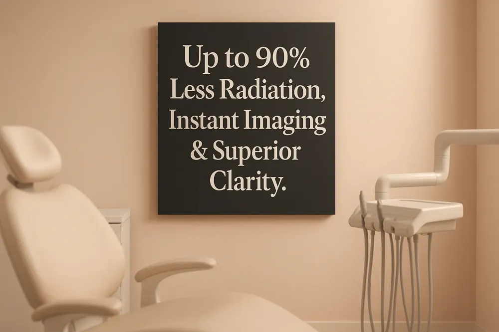
A primary advantage of digital systems is a significant reduction in radiation exposure. Digital dental X-rays use up to 90% less radiation compared to traditional film-based methods. For example, while a standard film X-ray might emit about 0.5 millirem, a digital X-ray can reduce this to as little as 0.1 millirem. This aligns perfectly with the ALARA principle—ensuring radiation doses are As Low As Reasonably Achievable—which guides all radiographic techniques in dentistry.
Speed is a hallmark of digital imaging. Instead of waiting for film to be chemically processed, images appear on a computer screen almost instantly after exposure. This immediate viewing drastically reduces the need for retakes, further minimizing patient exposure, and allows dentists to make quicker, more informed decisions. It also facilitates real-time collaboration with patients, visually explaining findings and discussing treatment options on the spot.
Digital technology provides clearer, more detailed images. With a wider dynamic range capturing up to 256 shades of grey—compared to about 25 in traditional X-rays—subtle details become visible. Dentists can digitally manipulate these images, adjusting contrast, brightness, and zoom, to spot tiny cavities, hairline fractures, and early signs of bone loss that might otherwise go undetected. This enhanced clarity supports early intervention, which can minimize the extent of future treatments.
| Comparison Aspect | Traditional Film Radiography | Modern Digital Radiography |
|---|---|---|
| Image Acquisition | Requires chemical development in a darkroom. | Instant display on a computer monitor. |
| Radiation Exposure | Higher dose required. | Up to 80-90% lower dose for the patient. |
| Image Manipulation | Not possible without retaking the X-ray. | Contrast, zoom, and filters can be applied. |
| Environmental Impact | Uses processing chemicals and physical film. | Eliminates chemical waste; uses electronic storage. |
| Record Management | Physical films require storage space. | Electronic files enable easy sharing and retrieval. |
Digital radiography is a foundational technology in modern dentistry that replaces traditional photographic film with electronic sensors to capture internal images of teeth and oral structures. Unlike conventional methods, it breaks the radiographic image into electronic data pieces for storage on a computer. This represents a major shift from the analog film processes used for decades. Since its introduction, digital radiography has become the standard in dental imaging due to its immediate benefits in safety, efficiency, and diagnostic capability.
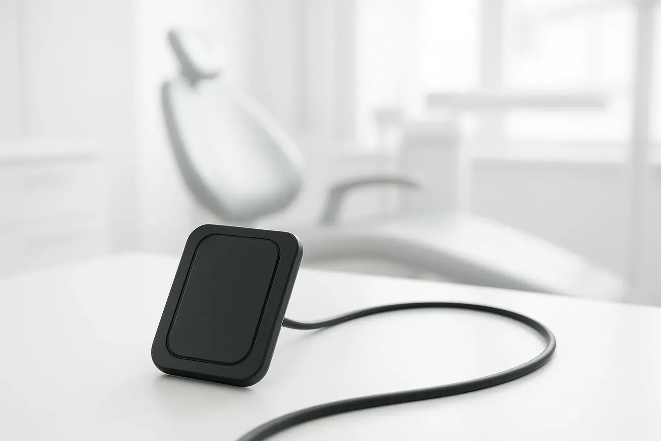
The process begins by placing a small, solid-state digital sensor inside the patient's mouth. When the X-ray machine is activated, the sensor captures the radiation energy that passes through the oral tissues. This captured energy is instantly converted into electronic signals. These signals are transmitted to a computer—often via a direct wire connection or, in modern wireless systems, via Bluetooth or Wi-Fi. Specialized imaging software then translates these signals into a high-resolution visual image that appears on a monitor within seconds.
Several sensor technologies are available, each with specific characteristics:
| Sensor Type | Full Name | Key Features & Usage |
|---|---|---|
| CCD | Charge-Coupled Device | One of the earliest digital sensors; provides direct, immediate image display. |
| CMOS | Complementary Metal Oxide Semiconductor | Common in modern sensors; offers efficient image capture efficiency and is often used in wireless systems. |
| PSP | Photo-Stimulable Phosphor | Uses a reusable phosphor plate that requires scanning after exposure; offers thin, flexible placement. |
Digital radiography entered dentistry with the introduction of the RadioVisioGraphy (RVG) launched in 1987, based on CCD and CMOS sensors. It gained widespread adoption through the late 1980s and 1990s, eventually overtaking conventional screen-film radiography. Today, it is an indispensable diagnostic tool used for detecting cavities, assessing bone health, planning treatments like implants, and monitoring oral health changes over time, forming a core component of contemporary patient-focused dental care.
| Covered Aspect | Core Explanation | Key Technological Detail |
|---|---|---|
| Core Definition | Replaces film with electronic sensors. | Images are digitized and stored on a computer. |
| Common Sensor Types | CCD or CMOS sensors and phosphor plates. | Differ in immediacy of image display and physical form. |
| Basic Process | Sensor capture, instant digitization, computer display. | Enables immediate viewing and image manipulation software. |
| Historical Introduction | Introduced to dentistry in 1987/1989. | Marked by the launch of the RVG system. |
Digital dental radiography offers a substantial reduction in radiation exposure compared to traditional film-based methods. Studies indicate a reduction of 50% to 90%, with intraoral digital radiography systems typically using 50%-60% less radiation than E-speed film. Some wireless digital sensors achieve an exposure reduction of up to 80%, delivering as little as 0.1 mrem per image versus 0.5 mrem for a standard film X-ray. This aligns with the core safety principle of ALARA (As Low As Reasonably Achievable), making digital imaging a significantly safer diagnostic tool.
The marked decrease in radiation dose is particularly crucial for children, pregnant individuals, and patients requiring frequent imaging, such as those undergoing orthodontic treatment or managing periodontal disease. For these groups, minimizing cumulative exposure is a primary concern. Digital radiography's ability to provide high-quality images with a fraction of the radiation addresses this need effectively, supporting safer long-term oral health management.
A defining feature of digital systems is instant image availability. Digital X-rays appear on a computer screen within seconds of exposure, eliminating the 10-15 minute wait required for film development. This immediacy allows dentists to review findings during the same appointment, facilitating quick and informed decisions and collaborative treatment discussions with the patient. It also drastically reduces the need for retakes due to processing errors, further minimizing unnecessary radiation exposure.
Digital X-rays provide superior diagnostic detail through high-resolution imaging and a wider dynamic range—capturing up to 256 shades of gray compared to about 25 in traditional film. This results in clearer pictures of teeth, bones, and soft tissues. Dentists can manipulate these digital images by adjusting contrast, brightness, and using software filters. They can also zoom in on specific areas to spot tiny cavities, early bone loss, or fractures that might be missed on conventional films, leading to more precise diagnosis.
Digital radiography eliminates the need for chemical processing, darkrooms, and physical film storage. This removes hazardous waste from the practice, reduces environmental pollution, and saves valuable office space. The workflow is streamlined as images are captured, stored, and retrieved electronically. Digital files can be easily shared with specialists or insurance companies via secure electronic transfer, enhancing care coordination and administrative efficiency.
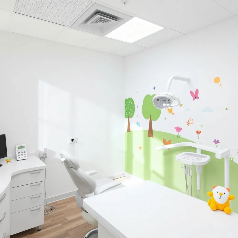
Digital sensors are typically thinner, smaller, and more flexible than traditional film packets. Wireless sensors, which use Bluetooth or Wi-Fi, offer even greater placement flexibility without cumbersome cables. This design improves comfort, especially for patients with sensitive gag reflexes or smaller mouths. The entire imaging procedure is faster, reducing the time a sensor needs to be held in position. While some studies note digital sensors can be less comfortable than film due to rigidity, the overall procedure time is shorter, contributing to a better patient experience.
| Feature | Digital X-Rays | Traditional Film X-Rays |
|---|---|---|
| Radiation Dose | Up to 90% less (typically 50%-80% reduction) | Higher, standard dose |
| Image Availability | Instant, on-screen viewing | Delay for chemical processing |
| Image Quality | High resolution, wide dynamic range, can be enhanced | Lower resolution, fixed contrast |
| Patient Comfort | Smaller, flexible sensors; faster procedure | Bulky film packets; longer placement time |
| Environmental Impact | No chemicals or film waste | Requires chemical processing and film disposal |
| Record Management | Easy electronic storage, retrieval, and sharing | Physical film storage, risk of degradation |

Modern digital dental X-rays use significantly less radiation than traditional film-based methods. The exposure from a single digital intraoral radiograph is extremely low, averaging about 0.005 millisieverts (mSv). This minute dose is a foundational element of the enhanced safety profile of contemporary digital imaging in dentistry.
To fully appreciate the minimal risk, it's helpful to compare this exposure to common sources of radiation. The average person in the United States receives about 3.2 mSv per year from natural background sources like radon gas, cosmic rays, and soil. A digital dental X-ray emits an amount of radiation comparable to just a few hours of this unavoidable background exposure.
| Common Radiation Source | Typical Dose (mSv) | Ratio Compared to Single Dental X-ray (~0.005 mSv) |
|---|---|---|
| Background Radiation (Annual) | 3.2 | 640 times greater |
| Cross-Country Flight | 0.04 | 8 times greater |
| Standard Chest X-ray | 0.1 | 20 times greater |
| Mammogram | 0.4 | 80 times greater |
| CT Scan (Abdomen & Pelvis) | 10 | 2000 times greater |
These comparisons underscore that the radiation dose from a necessary dental X-ray is negligible relative to exposures encountered daily and through other medical procedures.
All dental radiographic techniques adhere to the ALARA principle (As Low As Reasonably Achievable). This core safety guideline mandates using the lowest possible radiation dose to obtain diagnostically adequate images. It ensures that the significant diagnostic benefits of X-rays are achieved with minimal patient exposure.
Dental practices employ specific techniques and technologies to uphold the ALARA principle. Rectangular collimation limits the X-ray beam to the size of the sensor, protecting adjacent sensitive tissues like the eyes and thyroid.
Proper patient positioning and using equipment like receptor-holding devices ensure a high rate of success on the first attempt. This reduces the need for retakes, thereby minimizing any additional exposure for both patient and operator.
Despite the extremely low doses involved, lead aprons and thyroid collars are still commonly used. Their primary function is patient reassurance and comfort. Seeing these familiar safety measures helps alleviate anxiety.
Recent guidelines from the American Dental Association note that, due to the precision of modern digital X-ray machines, these shields are no longer mandated for all patients as the radiation risk is so small. However, many practices continue their use as a precaution, particularly for pregnant patients and children.
Yes, digital X-rays are generally considered safer than traditional film X-rays. They expose patients to significantly less radiation—often 50% to 90% less. This reduction in dose, combined with the elimination of chemical processing and immediate image viewing, represents a major advancement in patient safety and care efficiency.
There is no universal "safe" number of X-rays. Safety is determined by clinical necessity, not a specific count. Dentists prescribe dental X-rays based on a patient's individual oral health history, age, and symptoms, always following the ALARA principle.
For a healthy adult with low risk, routine bitewing X-rays might be recommended every 24 to 36 months. For patients with ongoing issues like gum disease or frequent decay, more frequent imaging may be necessary. The key is that the diagnostic benefit of detecting hidden problems far outweighs the minimal radiation risk involved with modern digital radiography.

The digital X-ray process is designed to be quick and comfortable for the patient. It begins with a consultation where the dentist reviews the patient's medical and dental history to determine the need for imaging. Before the procedure, patients are asked to remove jewelry or metal objects. For safety, protective gear such as a lead apron and thyroid collar may be used, though modern low-dose protocols mean these are sometimes optional.
Positioning is crucial for accuracy. The patient's head is stabilized, and the jaw and teeth are aligned with the machine. A small, thin digital sensor is then placed in the mouth. Modern wireless digital sensors are designed to be more comfortable and less bulky than traditional film packets. A controlled burst of radiation passes through the oral structures and is captured by the sensor. The image appears instantly on a computer monitor, eliminating the waiting time associated with film development.
Once captured, the digital image is processed and enhanced using specialized software. This capability is a significant diagnostic advantage. Dentists can adjust contrast and density to bring out specific details. Specialized software filters, such as caries or periodontal filters, can highlight structures relevant to specific conditions.
For instance, contrast enhancement can make subtle bone changes more visible. Zooming in on a high-resolution image allows for detailed examination of a tiny fracture or early decay. Digital Subtraction Radiography (DSR) software can precisely compare images taken at different times to detect minute changes, such as early bone loss in periodontal disease or the progression of a cavity. These manipulations provide more information without exposing the patient to additional radiation.
Immediate image availability transforms patient consultations. Dentists can display the enhanced images on a chairside monitor and explain findings visually. Patients can see areas of concern, such as the start of decay between teeth or bone levels around a tooth, making the diagnosis clear and tangible.
This visual aid builds trust and facilitates a collaborative approach to treatment planning. Patients become active participants, better understanding their condition and the proposed treatments. Real-time discussions about treatment options, based on clear visual evidence, lead to more informed decisions and higher patient engagement in their own oral healthcare.
Digital radiography streamlines practice administration and enhances continuity of care. Images are stored electronically in a patient's digital chart, often in secure cloud-based systems. This eliminates physical film storage, saves office space, and reduces environmental waste from chemical processing. Electronic records are easy to retrieve for future appointments, enabling easy comparison of a patient's oral health over time.
Digital files can be securely and instantly shared with specialists for consultations or with insurance companies to expedite claims. The use of the universal DICOM (Digital Imaging and Communications in Medicine) standard ensures compatibility across different systems. This seamless digital workflow supports efficient record-keeping and improves coordination among all dental professionals involved in a patient's care.
| Process Stage | Key Activities & Features | Direct Patient & Practice Benefits |
|---|---|---|
| Procedure | Consultation, sensor placement, quick image capture. | Faster appointment, enhanced patient comfort, immediate results. |
| Diagnosis | Software enhancement, zoom, contrast/filter application. | Higher accuracy, no extra radiation for adjustments. |
| Consultation | On-screen image viewing and explanation. | Better understanding, collaborative treatment planning. |
| Administration | Electronic storage, secure sharing, digital records. | Easy retrieval, efficient insurance claims, specialist collaboration. |

Digital radiography provides faster image acquisition and viewing, eliminating the wait for film development. It reduces patient radiation exposure by up to 90% lesser radiation dose compared to traditional film. The technology offers superior image quality, allowing for digital enhancement through software filters without additional exposure. Images are stored electronically on a computer system, facilitating easy retrieval, sharing with specialists, and tracking of oral health changes over time. The process is also more environmentally friendly, eliminating chemical developers.
Disadvantages include higher initial equipment costs and the potential fragility of some wireless digital sensors. Some patients may find certain intraoral sensors bulky compared to film. While image quality is generally high, some studies have indicated conventional film superiority in certain subjective image quality comparisons, though modern digital systems continue to improve.
Digital dental X-rays are indispensable for diagnosing conditions not visible during a routine visual exam. Their key applications include:
Dental X-rays are broadly categorized as intraoral (taken inside the mouth) and extraoral (taken outside the mouth).
Intraoral X-rays provide exceptionally detailed images and are the most frequently used.
| Type | Area Captured | Primary Diagnostic Use |
|---|---|---|
| Bitewing | Crowns of upper and lower teeth in a specific area. | Detecting decay between teeth and monitoring bone health. |
| Periapical | Entire tooth from crown to root tip and surrounding bone. | Diagnosing root infections, abscesses, and bone changes. |
| Occlusal | Full arch of teeth in either the upper or lower jaw. | Viewing tooth development, fractures, or abnormalities in the jaw's floor or roof. |
Extraoral X-rays provide a broader view of the skull and jaws.
| Type | Area Captured | Primary Diagnostic Use |
|---|---|---|
| Panoramic | All teeth, jaw joints, sinuses, and nerves in a single 2D image. | General screening, orthodontic planning, wisdom tooth assessment. |
| Cephalometric | Side view of the entire head. | Orthodontic treatment planning to analyze jaw and tooth relationships. |
For complex cases, Cone Beam CT provides detailed 3D dental imaging. It is invaluable for planning dental implants, evaluating jaw pathology, and assessing intricate root canal anatomy. CBCT uses a higher radiation dose than standard 2D digital X-rays, so its use is carefully justified based on patient history and clinical need where conventional radiographs provide inadequate information.
Digital X-rays are a powerful tool but are only one part of a comprehensive diagnostic assessment. Their use is always justified by the patient's specific symptoms, health history, and clinical examination findings. Dentists adhere to the ALARA principle to minimizing radiation exposure, using techniques like faster digital sensors and rectangular collimation. For many patients, routine check-up X-rays are recommended every 6 to 18 months, though frequency depends on individual oral health risks.
| Technology | Key Feature | Common Applications | Key Limitation / Note |
|---|---|---|---|
| Digital Radiography | 2D imaging; immediate viewing; lower radiation doses. | Routine exams, cavity detection, bone assessment. | Standard diagnostic workhorse for dentistry. |
| Panoramic | Broad single-image view of jaws and teeth. | Screening, ortho planning, wisdom teeth. | Lower resolution than intraoral views. |
| Cone Beam CT (CBCT) | Detailed 3D imaging of bone and teeth. | Implant planning, complex surgery, endodontics. | Higher radiation dose; not for routine screening. |
The integration of digital radiography represents a fundamental shift towards more gentle and patient-focused dentistry. By leveraging advanced sensor technology, it removes the need for cumbersome film and chemical processing, streamlining the entire imaging experience. This approach prioritizes patient comfort and convenience, embodying the modern standard for dental care.
A primary advantage of digital systems is a significant reduction in radiation exposure—up to 90% less than traditional film-based methods. Adhering to the ALARA (As Low As Reasonably Achievable) principle, this technology minimizes patient risk. This makes it a safer choice for all patients, including children and those requiring frequent imaging. The radiation dose from a single digital dental X-ray is remarkably low, comparable to just a few hours of natural background exposure.
Digital radiography provides immediate results. Images appear on-screen within seconds, eliminating the waiting period required for film development. This rapid acquisition enables quicker diagnosis and allows dentists to discuss findings with patients in real-time. The efficiency reduces appointment times and facilitates prompt treatment planning, enhancing the overall patient experience.
The high-resolution images produced by digital sensors offer superior detail and can be manipulated without additional radiation exposure. Dentists can adjust contrast, zoom in on specific areas, and apply software filters. This enhanced clarity allows for the detection of subtle issues, such as early decay between teeth, minor fractures, and initial bone loss, that might be missed with traditional radiographs.
The combination of speed, safety, and image quality makes digital radiography an invaluable tool for early intervention. By identifying problems at their inception, treatments can be less invasive and more conservative. The ability to store and compare digital images over time also supports the monitoring of oral health changes, enabling truly personalized and proactive treatment plans tailored to each patient's unique needs.
Embracing digital radiography reflects a commitment to utilizing the most advanced technology available for optimal patient outcomes. It supports a collaborative approach to dental health, where clear, enhanced images foster better understanding and informed decision-making between the dentist and patient. This technology is not merely an upgrade in equipment but a cornerstone of a modern practice dedicated to safety, precision, and superior diagnostic care.
| Aspect of Care | Digital Radiography Contribution | Patient Benefit |
|---|---|---|
| Safety Profile | Reduces radiation dose by up to 90% | Minimizes exposure, especially for vulnerable groups |
| Diagnostic Speed | Images available instantly for review | Enables faster diagnosis and treatment discussions |
| Image Utility | High-resolution, digitally manipulable files | Allows for detailed examination and early problem detection |
| Record Management | Electronic storage and easy retrieval | Simplifies tracking of oral health changes over time |
| Environmental Impact | Eliminates chemical processing and film waste | Supports eco-friendly practice operations |